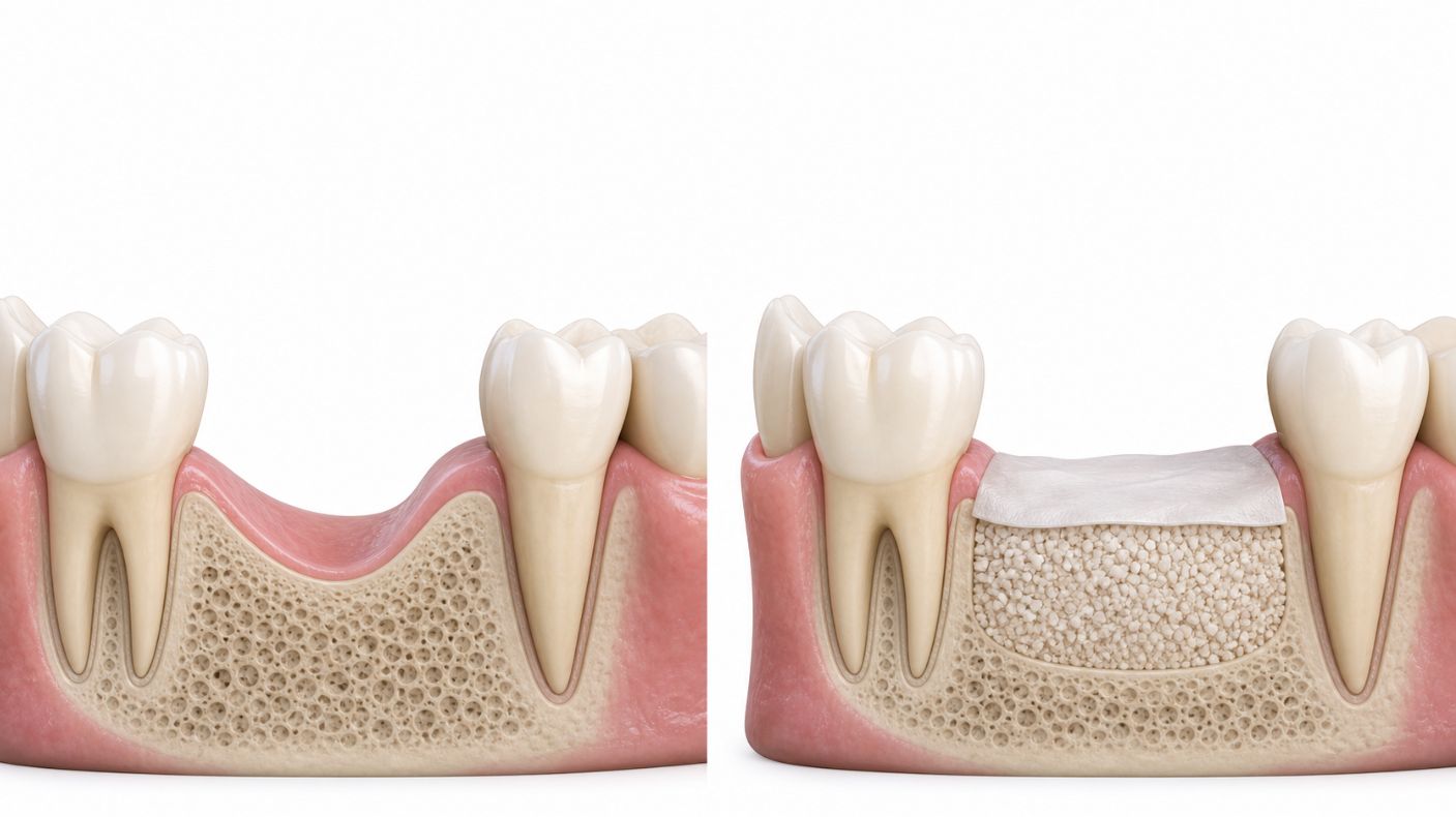
When a tooth is removed, the empty space left behind is not simply a gap in the gum—it is a healing bone wound. In the first three to six months after extraction, the jawbone around that socket naturally changes shape and volume. This process can make future dental implant treatment more complex, less predictable and, in some cases, more expensive.
Bone socket preservation, also called ridge preservation, alveolar ridge preservation or socket grafting, is a procedure performed at the time of extraction to reduce this post-extraction bone loss. It usually involves placing a bone graft material into the socket and protecting it with a collagen membrane, plug or sutures. The aim is not to “freeze” the jawbone exactly as it is, but to preserve as much height, width and natural contour as reasonably possible for a future dental implant, bridge or denture.
For patients considering an implant after tooth removal, the key discussion should happen before the extraction—not months later, when the ridge may already have narrowed.
Important: This article provides general UK patient education and is not a substitute for an examination, X-rays or a personalised treatment plan from a dentist, oral surgeon or periodontist.
What Is Bone Socket Preservation?
Bone socket preservation is a preventive dental procedure performed immediately after, or sometimes shortly after, a tooth extraction. Once the tooth has been gently removed, the clinician cleans the socket, assesses the remaining bone walls and places a grafting material into the site where the roots once sat.
The material may be covered with a collagen membrane, collagen sponge, platelet-rich fibrin, or the patient’s own gum tissue. Fine sutures may be used to secure the protective covering. Over the following months, the body heals the socket and gradually remodels the graft material alongside newly forming bone.
The terminology can sound confusing, but these terms generally refer to the same treatment objective:
The procedure is most often considered where a future implant is planned but cannot be placed immediately, where the visible front-tooth area is involved, where bone walls are thin or damaged, or where an extraction is expected to leave a significant defect.
Why Does the Jawbone Shrink After an Extraction?
The bone that surrounds and supports teeth is called the alveolar bone. It exists largely because teeth and their roots are present. When a tooth is removed, the body no longer receives the same mechanical stimulation in that area, and the supporting bone begins to remodel.
This is a normal biological response, not a sign that something has gone wrong. The extraction socket initially fills with a blood clot. The clot is then replaced by healing tissue, immature bone and eventually more mature bone. At the same time, however, the outer walls of the ridge – particularly the thin facial or cheek-side bone – may resorb.
The cheek-side bone wall is often naturally thinner than the bone on the tongue or palate side. It is therefore especially vulnerable to post-extraction loss. This is clinically important in the upper front teeth, where small changes in bone and gum contour can affect the appearance of a future implant crown.
Without ridge preservation, healing may result in a ridge that is lower, narrower and shifted slightly towards the palate or tongue. The gum tissue follows the underlying bone, meaning the visual contour of the smile can also change.
A major systematic review found that, by around six months after extraction, horizontal bone reduction can range from 29% to 63%, while vertical reduction can range from 11% to 22%. On average, horizontal loss was greater than vertical loss.
What Happens During the First 3 to 6 Months?
The most significant remodelling usually happens early. Although the exact pattern differs between patients and extraction sites, the following timeline helps explain why immediate planning matters.
First 24 Hours: Blood Clot Formation
Immediately after a tooth is extracted, bleeding fills the socket and a blood clot forms. This clot is the body’s first protective dressing. It covers exposed bone and nerve endings and creates the foundation for normal healing.
The early clot is fragile. Patients are usually advised not to smoke, vape, rinse forcefully, spit aggressively or drink through a straw during the first day because these actions may disturb it. If the clot breaks down prematurely, a painful complication called dry socket can develop.
For ridge preservation, the clinician uses this early healing opportunity to place graft material into the clean socket before the bone begins substantial collapse.
First 1 to 2 Weeks: Soft Tissue Healing Begins
During the first week or two, the blood clot is gradually replaced by granulation tissue. This is healing tissue containing small blood vessels, cells and collagen. The gum edges begin closing over the socket.
If a collagen membrane or plug has been placed as part of socket preservation, it helps protect the graft and acts as a barrier during this delicate phase. Some membranes dissolve naturally; others require removal, depending on the material and technique used.
It is normal to have mild swelling, bruising, tenderness or occasional small graft particles appearing in the mouth during early healing. However, severe pain, increasing swelling, persistent bleeding, fever or a bad taste should be reported to the treating dentist promptly.
First 4 to 8 Weeks: Early Bone Formation
As healing progresses, new woven bone starts forming inside the socket. The socket may look closed at the surface, but the deeper bone is still immature and actively remodelling.
This is an important point for patients: gum closure does not mean that the bone underneath has completed healing. The socket may appear healed in a mirror after several weeks, while biological remodelling continues beneath the surface for months.
In a non-grafted socket, ridge reduction is already under way. The body resorbs and reshapes the bone walls, especially where the facial wall was thin or damaged before extraction.
Around 3 Months: Noticeable Ridge Remodelling
By three months, much of the rapid early change has already occurred. Research has reported approximately 32% horizontal ridge reduction at this stage in untreated extraction sites.
This loss is important because implants need adequate bone around them for stability, function and appearance. If the ridge becomes too narrow, an implant cannot simply be placed in the centre of the original socket without risking poor positioning, inadequate bone coverage or an unnatural crown shape.
At this stage, some patients may be suitable for implant placement, depending on the original site, the graft material used, the quality of healing and the clinician’s assessment. Others need a longer healing period.
Around 4 to 6 Months: Maturing Bone and Implant Planning
At four to six months, the socket graft is generally more mature and the site can be assessed for the next stage of treatment. A dentist may use a clinical examination, periapical X-ray, panoramic X-ray or CBCT scan to evaluate bone volume and plan implant position.
The ideal timing depends on the individual case. Some clinical guidance and reviews describe delayed implant placement at approximately four to six months following socket preservation, while other protocols may require longer healing.nature+1
By six months, the difference between a preserved ridge and an untreated ridge can be significant. A systematic review found that grafting a socket can reduce the amount of bone resorption when compared with extraction alone, including in horizontal and vertical dimensions.
Why Leaving an Empty Socket Can Complicate Implant Treatment
An implant needs more than a hole in the jawbone. It needs adequate bone in the right three-dimensional position: enough width from cheek to tongue, enough height, healthy surrounding gum and a position that allows the final crown to look and function like a natural tooth.
When an extraction socket heals without preservation, several challenges can arise.
A Narrow Ridge May Not Fit an Implant Properly
Modern dental implants are small, but they still require sufficient bone around them. If the ridge loses width, the clinician may not be able to place an implant in the ideal position without further grafting.
An implant placed too far towards the palate or tongue may be difficult to restore naturally. The crown may need to be over-contoured, harder to clean or less aesthetically pleasing. In the front of the mouth, even a small positional compromise can affect the smile line.
Bone Height May Also Reduce
Vertical bone reduction can make treatment more difficult, particularly near important anatomical structures. In the upper back jaw, reduced height may bring the maxillary sinus closer to the implant site. In the lower back jaw, bone loss may reduce the available distance from the inferior alveolar nerve.
These situations do not necessarily prevent implant treatment, but they can require additional procedures such as sinus augmentation, guided bone regeneration or a different implant design.
The Gum Contour May Flatten
Bone and gum are closely connected. When bone volume reduces, the overlying soft tissue may flatten or recede. In visible areas, this can result in a sunken appearance above an implant crown or bridge pontic.
Preserving ridge contour is particularly relevant for upper front teeth, high smile lines, thin gum tissue and patients seeking highly aesthetic implant treatment. Socket preservation cannot guarantee a perfect cosmetic result, but it gives the clinician a better starting point.
Later Grafting Can Be More Invasive
If bone loss has already occurred, a patient may still be able to receive an implant. However, they may need a larger or more complex grafting procedure later.
Delayed augmentation can involve opening the gum, adding graft material to the outside of the ridge, using a barrier membrane, securing the graft with pins or screws, and allowing several additional months for healing. These treatments can be highly successful, but they may increase cost, treatment time and surgical complexity.
Socket preservation is therefore often a risk-reduction strategy: a relatively contained procedure performed at the time of extraction to reduce the likelihood of more extensive treatment later.
Does Everyone Need a Bone Graft After Extraction?
No. Socket preservation is not necessary after every tooth extraction, and it should not be presented as an automatic add-on without a clinical reason.
A dentist may decide that a socket can heal naturally when:
-
No implant or fixed replacement is planned
-
The patient will use a removable denture and adequate ridge volume is present
-
The socket has thick, intact bone walls
-
An implant can be placed immediately and securely at the same appointment
-
Medical, financial or treatment-preference factors make grafting unsuitable
-
The clinician determines that future bone volume is unlikely to be a limiting issue
However, ridge preservation deserves serious consideration in certain situations:
-
A dental implant is planned after healing
-
The implant will be delayed for several months
-
The tooth is in the visible smile zone
-
The cheek-side bone wall is thin, missing or damaged
-
There is existing gum disease, bone loss or infection-related damage
-
Several adjacent teeth are being removed
-
The patient has thin gum tissue or a high smile line
-
Maintaining ridge shape is important for a bridge pontic or denture
The decision should be based on a proper assessment, not a one-size-fits-all rule. Your clinician may take X-rays or a CBCT scan and discuss whether immediate implant placement, socket preservation, delayed implant placement or a non-implant replacement is the most sensible route.
What Materials Are Used for Socket Grafting?
There is no single “best” graft material for every case. The choice depends on the socket anatomy, whether the bone walls are intact, the planned restoration, clinician preference, patient values and the desired healing timeline.
Common material categories include:
Patients should feel comfortable asking exactly what material is proposed and why. This can be particularly important for people with religious, ethical, dietary or personal concerns about animal-derived or human donor materials.
A collagen membrane or plug may also be used. Its role is to protect the graft, reduce soft-tissue invasion into the socket and support guided bone regeneration. The evidence does not establish one universally superior material or barrier for every patient and every socket.
What Does the Procedure Involve?
Socket preservation is usually carried out under local anaesthetic as part of the extraction appointment.
A typical process includes:
-
The dentist or oral surgeon removes the tooth as gently as possible to preserve the surrounding bone walls.
-
The socket is inspected and carefully cleaned of infection, inflamed tissue or debris.
-
The clinician evaluates whether the socket walls are intact and whether additional treatment is needed.
-
Bone graft particles are placed into the socket without excessive compression.
-
A collagen plug, membrane or soft-tissue graft is positioned over the area.
-
Sutures may secure the site.
-
The patient receives aftercare instructions and a review appointment.
The procedure may add time and cost to the extraction visit, but it can avoid or simplify later grafting. Most patients experience a recovery similar to that after a surgical extraction, although discomfort varies according to the tooth, procedure complexity and individual healing.
Benefits and Limitations of Ridge Preservation
The main benefit is maintaining more bone volume than would be expected with extraction alone. Meta-analysis data suggest ridge preservation can reduce loss in both ridge width and height, although the amount of benefit varies between studies and techniques.
A 2021 Cochrane review found that ridge-preservation methods may reduce post-extraction dimensional changes, but judged the certainty of the evidence to be very low and noted limited long-term evidence regarding implant failure, aesthetic outcomes and the need for later augmentation.
That means patients should receive balanced advice. Socket preservation is not a guarantee that no further grafting will ever be needed. It does not create unlimited bone, correct every pre-existing defect or guarantee an implant. Instead, it can make future treatment more feasible and preserve options.
Potential limitations and risks include:
-
Cost at the extraction appointment
-
Temporary swelling, bruising or discomfort
-
Small graft particles becoming loose during healing
-
Membrane exposure or delayed healing
-
Infection, although uncommon with good surgical technique and aftercare
-
The possibility that further grafting is still required before implant placement
-
A longer overall treatment timeline before the final implant crown
UK Aftercare Following Socket Preservation
Your own dentist’s instructions always take priority, but UK post-operative advice commonly includes protecting the blood clot and graft during early healing.
For the first 24 hours, avoid rinsing, forceful spitting, smoking, vaping, alcohol and strenuous exercise. Eat soft foods once the numbness has worn off, and avoid chewing directly on the treated side.
From the following day, gentle warm salt-water rinses may be recommended. Brush the rest of your teeth normally but take care around the surgical site. Do not pull at stitches, probe the socket or repeatedly check it with your tongue.
Smoking and vaping are particularly important to avoid because nicotine and suction can compromise blood supply and wound stability. If you are finding this difficult, speak to your dentist, pharmacist or GP about stop-smoking support.
Call your dentist or oral surgery provider if you experience worsening rather than improving pain, persistent bleeding, increasing swelling, fever, pus, a foul taste or a membrane that has moved substantially.
Questions to Ask Before an Extraction
Before consenting to an extraction, consider asking:
-
Am I likely to want a dental implant in this space later?
-
How much bone support is currently present around this tooth?
-
Is the cheek-side bone wall thin or damaged?
-
Would socket preservation improve my implant options?
-
Can an implant be placed immediately, or should it be delayed?
-
What graft and membrane materials are being recommended?
-
Are the materials animal-derived, donor-derived or synthetic?
-
How long should I wait before implant planning?
-
What happens if I choose extraction without grafting?
-
What are the likely costs now compared with possible delayed grafting later?
A good consultation should make the options clear without pressure. In some cases, allowing the socket to heal naturally is entirely reasonable. In others, preserving the ridge at the extraction appointment may be the most cost-effective and biologically sensible step towards a future implant.
Conclusion
Tooth extraction starts a predictable process of jawbone remodelling. The greatest change often occurs in the first three to six months, with untreated sites commonly losing more width than height. This can affect not just implant placement, but also gum contour, bridge design, denture stability and the long-term appearance of the smile.
Bone socket preservation is a proactive treatment that aims to limit—not eliminate—this natural bone loss. For patients considering a future implant, the best time to discuss it is before the tooth is removed. A personalised assessment by a UK dentist, periodontist or oral surgeon can determine whether immediate bone grafting is appropriate for your extraction site and future goals.
Bone Socket Preservation FAQs
What is bone socket preservation?
Bone socket preservation, also called ridge preservation or socket grafting, is a procedure carried out after tooth extraction to reduce the natural loss of jawbone volume. A dentist places graft material in the empty socket, often with a protective collagen membrane or plug, to support healing and maintain the ridge shape for a future implant, bridge or denture.
Do I need a bone graft after every extraction?
No. Bone grafting is not necessary after every tooth removal. It is most often recommended when you may want a dental implant later, when the tooth is in a visible area, when the socket walls are thin or damaged, or when significant bone shrinkage is expected.
Why does bone shrink after a tooth is removed?
The jawbone around tooth roots is called alveolar bone. Once the root is removed, the body naturally remodels this bone because it no longer has the same function or stimulation. The ridge can lose both width and height, with the cheek-side bone often particularly vulnerable.pubmed.
How much bone loss happens after an extraction?
Bone loss varies according to the tooth, socket anatomy, gum health and healing factors. Research has reported horizontal ridge reduction of about 32% after three months and 29% to 63% at six to seven months in untreated extraction sites; vertical loss has been reported at around 11% to 22% after six months.
When is the best time for socket preservation?
The ideal time is usually at the same appointment as the extraction. Grafting a fresh socket gives the dentist an opportunity to support the ridge before substantial post-extraction resorption begins.
Can I have an implant placed immediately instead?
Sometimes. Immediate implant placement may be suitable where there is enough healthy bone to achieve stability, no uncontrolled infection and a favourable bite and gum situation. It is not suitable for every case, and your dentist may recommend ridge preservation with delayed implant placement instead.
Does socket preservation guarantee that I will not need another graft?
No. Socket preservation reduces bone loss; it does not stop all natural remodelling or guarantee that an implant can be placed without further grafting. It can, however, preserve more bone volume and may reduce the scale of any future augmentation required.onlinelibrary.
What materials are used for socket grafting?
The graft may be synthetic, animal-derived, processed human donor bone or bone taken from another area of your mouth. A collagen membrane or plug may cover the graft. Ask your dentist what material is being used, whether it is animal- or donor-derived, and why it is suitable for your case.
Is socket preservation painful?
The procedure is normally completed with local anaesthetic, so you should not feel pain during treatment. Afterwards, soreness, mild swelling and bruising are possible, particularly if the extraction itself was surgical. Your dental team will give you specific pain-relief and aftercare instructions.
How long does it take for a socket graft to heal?
Soft tissue may look healed within a few weeks, but the bone beneath continues to mature for months. Implant planning commonly takes place around four to six months after preservation, although the exact timing depends on the graft material, extraction site and your clinician’s assessment.
Can I smoke or vape after a socket graft?
It is best to avoid smoking and vaping during healing. Nicotine can affect blood supply and wound repair, while suction from inhaling can disturb the healing site. Follow your dentist’s specific advice on how long to avoid these products.
What should I eat after socket preservation?
Choose soft, lukewarm or cool foods initially, such as yoghurt, scrambled eggs, soup that is not hot, mashed potato or pasta. Avoid chewing directly on the treated area, and avoid hard, sharp or crumbly foods that could disturb the site or become trapped around the graft.
Can graft particles come out after surgery?
A small number of loose graft granules can occasionally be noticed during early healing. This is not always a problem, but contact your dentist if a large amount of material is lost, the membrane moves, bleeding continues, or pain and swelling worsen.
What happens if I do not have socket preservation?
The socket will still heal naturally, but the surrounding ridge is likely to shrink to some extent. If you later decide to have an implant, you may need additional bone grafting, a longer treatment timeline or a different tooth-replacement option.
Is ridge preservation safe?
Socket preservation is a widely used procedure, but every surgical treatment has risks. Possible complications include infection, swelling, membrane exposure, graft loss, delayed healing and the need for further grafting. A dentist can explain your individual risks after examining the tooth and reviewing your medical history.
When should I contact my dentist after socket grafting?
Contact your dentist promptly if you have severe or worsening pain, increasing swelling, persistent bleeding, fever, pus, a foul taste, a loose membrane or extensive graft loss. Seek urgent medical help if you develop difficulty breathing or swallowing.
Sources
- Tan et al.: A systematic review of post-extraction alveolar hard and soft tissue dimensional changes
- Cochrane Review: Alveolar ridge preservation techniques for dental implant site development
- British Dental Journal: Socket preservation
- Systematic review: Effect of alveolar ridge preservation after tooth extraction
- Socket preservation and guided bone regeneration review
- Extraction socket preservation: Principles and indications